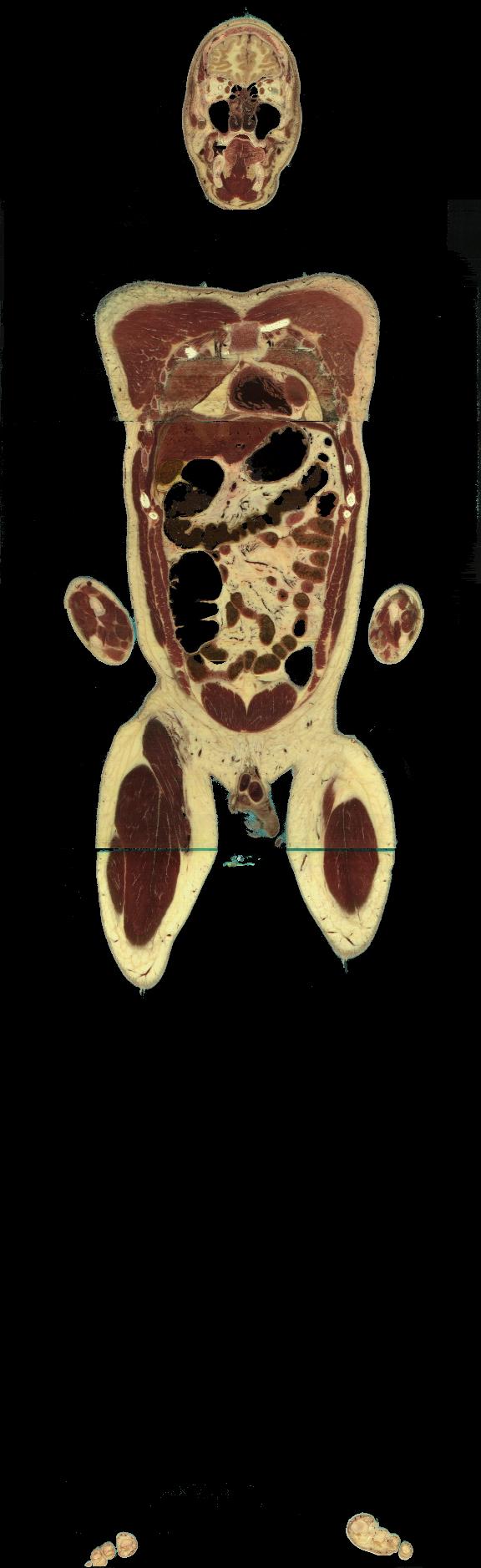
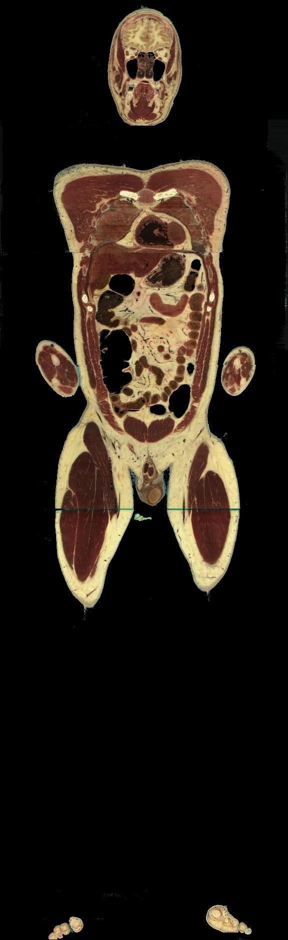
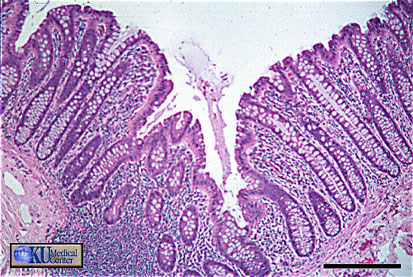
The colon has no pitts or villi; divided into cecum, appendix, ascending, transverse, descending, and sigmoid colons, rectum, and anal canal. Functions in absorption of water, electrolytes, some vitamins, remaining amino acids, lipids, and carbohydrates; compacts feces.
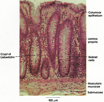
MUCOSA (tunica mucosa):
- epithelium (wet surface epithelia): simple (surface epithelial cell) columnar absorptive epithelium with abundant goblet (oligomucous) cells,
- lamina propria: underlying loose ct; glands (= crypts of Lieberkühn)m simple columnar epithelium, regenerative cells, and APUD (enteroendocrinocytes) cells in base release paracrine hormones
- muscularis mucosae: thin layer smooth muscle
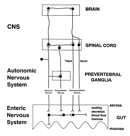
SUBMUCOSA: regular/irregular (coarse) fibroelastic ct; submucosal (Meissners) nervous plexuses (pre- and post-ganglionic parasympathetic fibers; nonmyelinated preganglionic fibers from vagus).
MUSCULARIS EXTERNA: two muscle layers (inner circular [tight helix; modified in anal sphincters]; outer longitudinal [loose helix]) modified as taniae coli: 3 thickening separate haustra coli (Roman device for hauling water; sacculations); myenteric (Auberbachs) nervous plexuses; sympathetic ganglia and fibers between muscle layers; peristaltic action independent;
SEROSA (ADVENTITIA): irregular dense ct surrounded by mesothelium (serosa) or bound to body wall (adventitia); appendices epiploicae = small fat-filled pouches
Appendix: surface epithelium with many goblet cells; glands relatively shallow; lamina propria infiltrated with lymphoid cells; lymph nodules in submucosa
Anorectal junction: abrupt change at anal valves from simple columnar of rectum to stratified squameous epithelium (keratinizing type) of anal canal; rectal glands short; lamina propria infiltrated by lmypohoid cells.
Anal Canal: anal columns = longitudinal folds joined at orifice to form anal valves and anal sinuses. Circumanal glands, hair follicles, and sebaceous glands. Spinincters formed by muscularis externa.